
- Home
- Projects
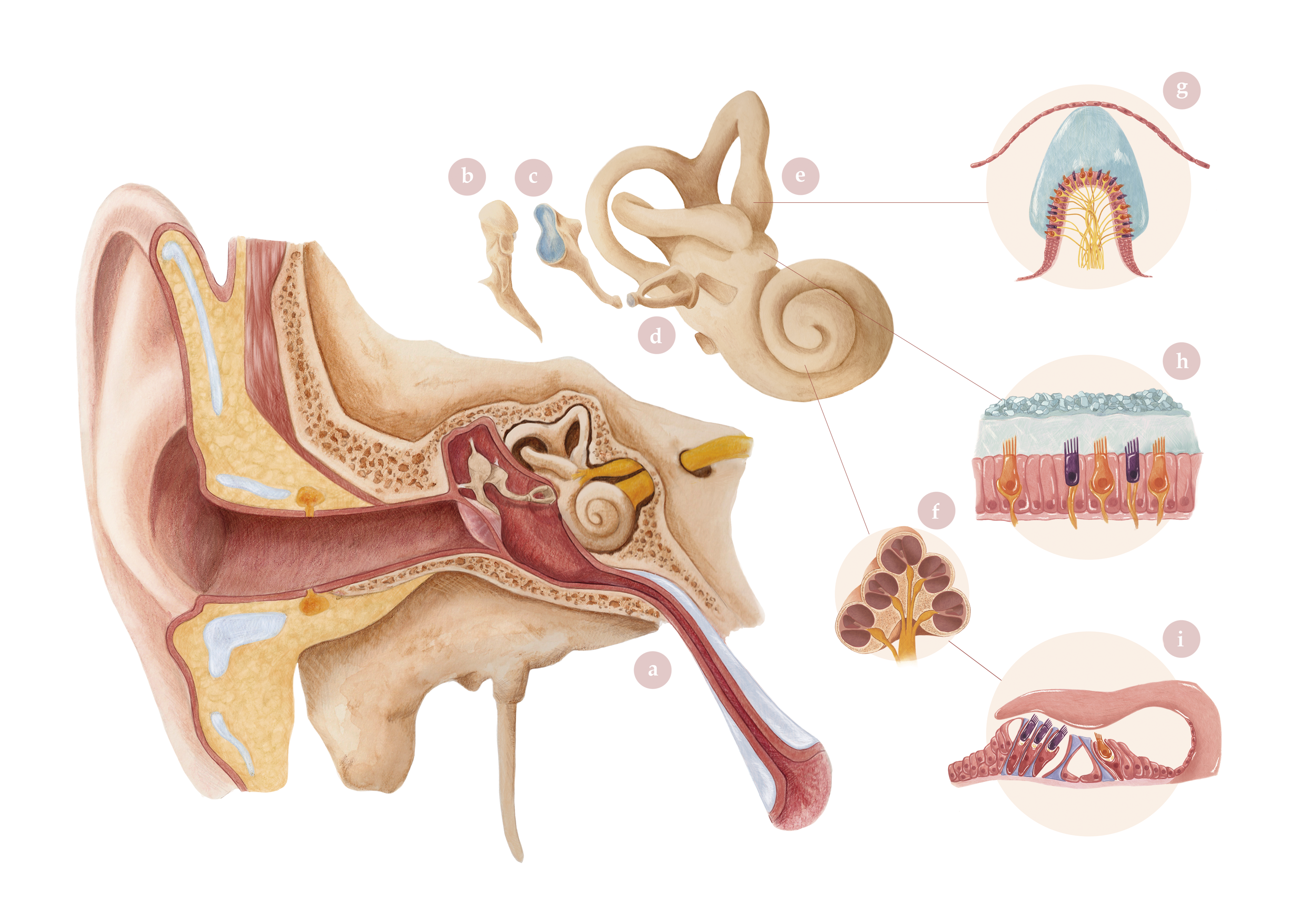
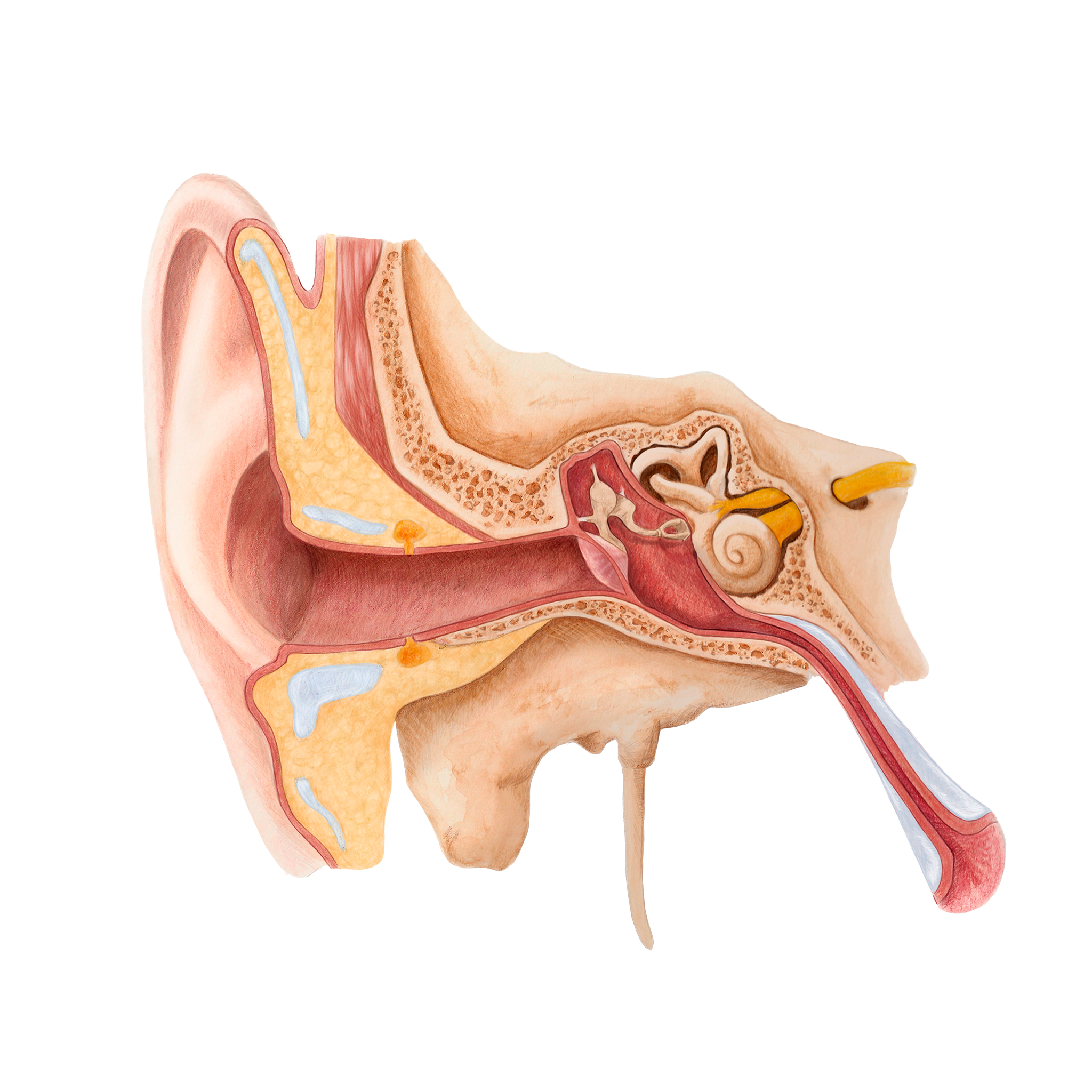
Anatomy of the ear and associated sensory organs
- Tipo de proyecto: Scientific illustrationType of project:
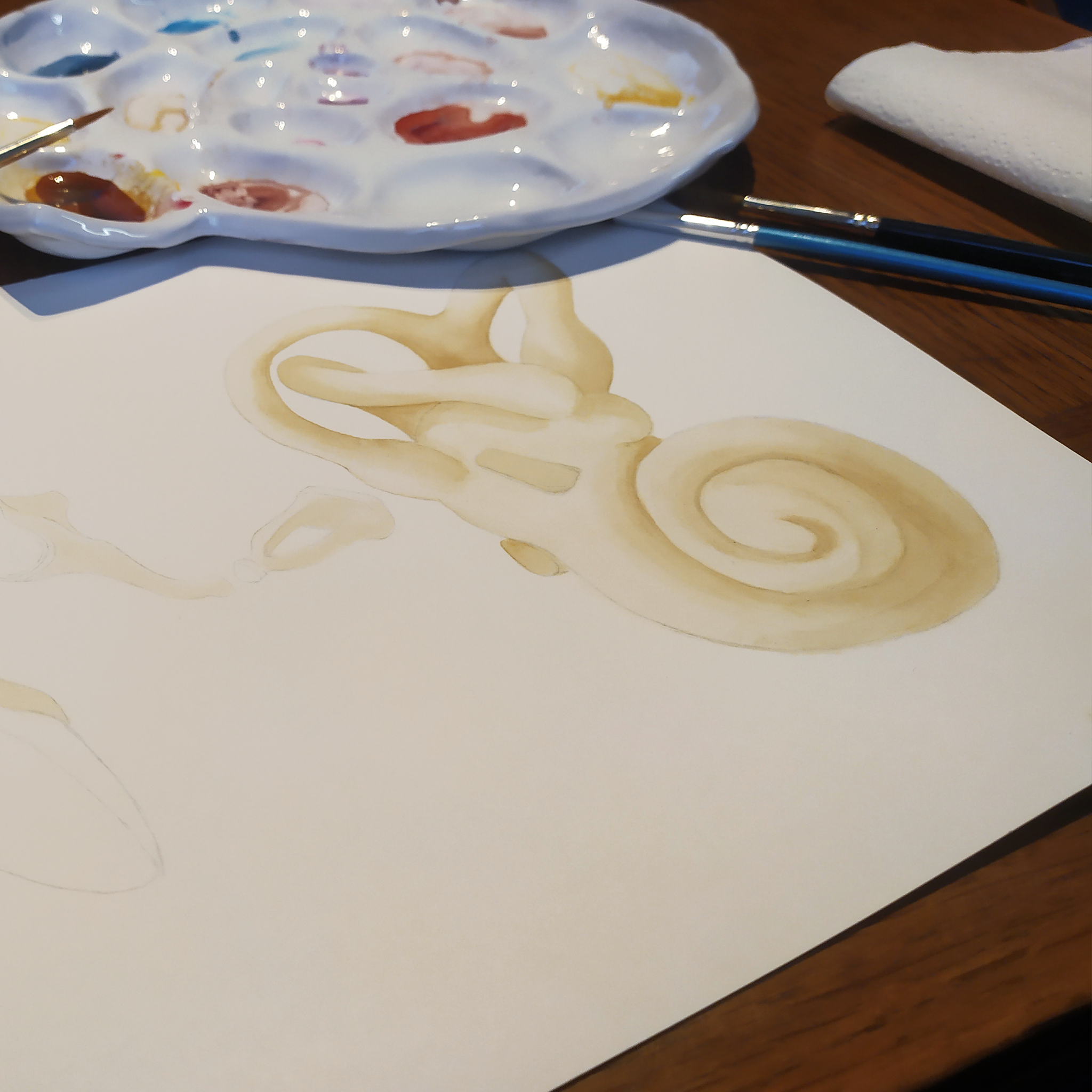
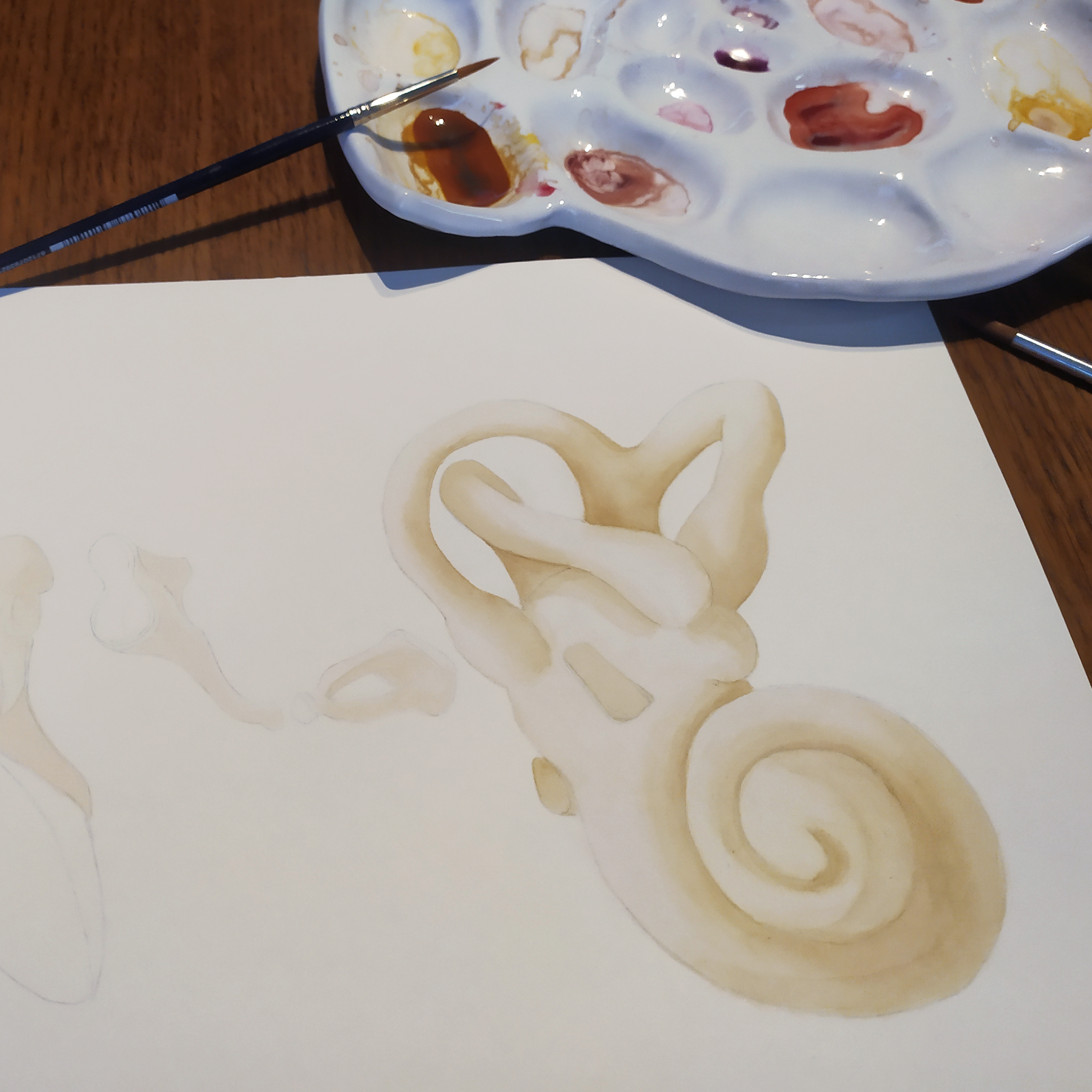
- Técnica: Mixed media: Watercolor, colored pencils, and digital illustration
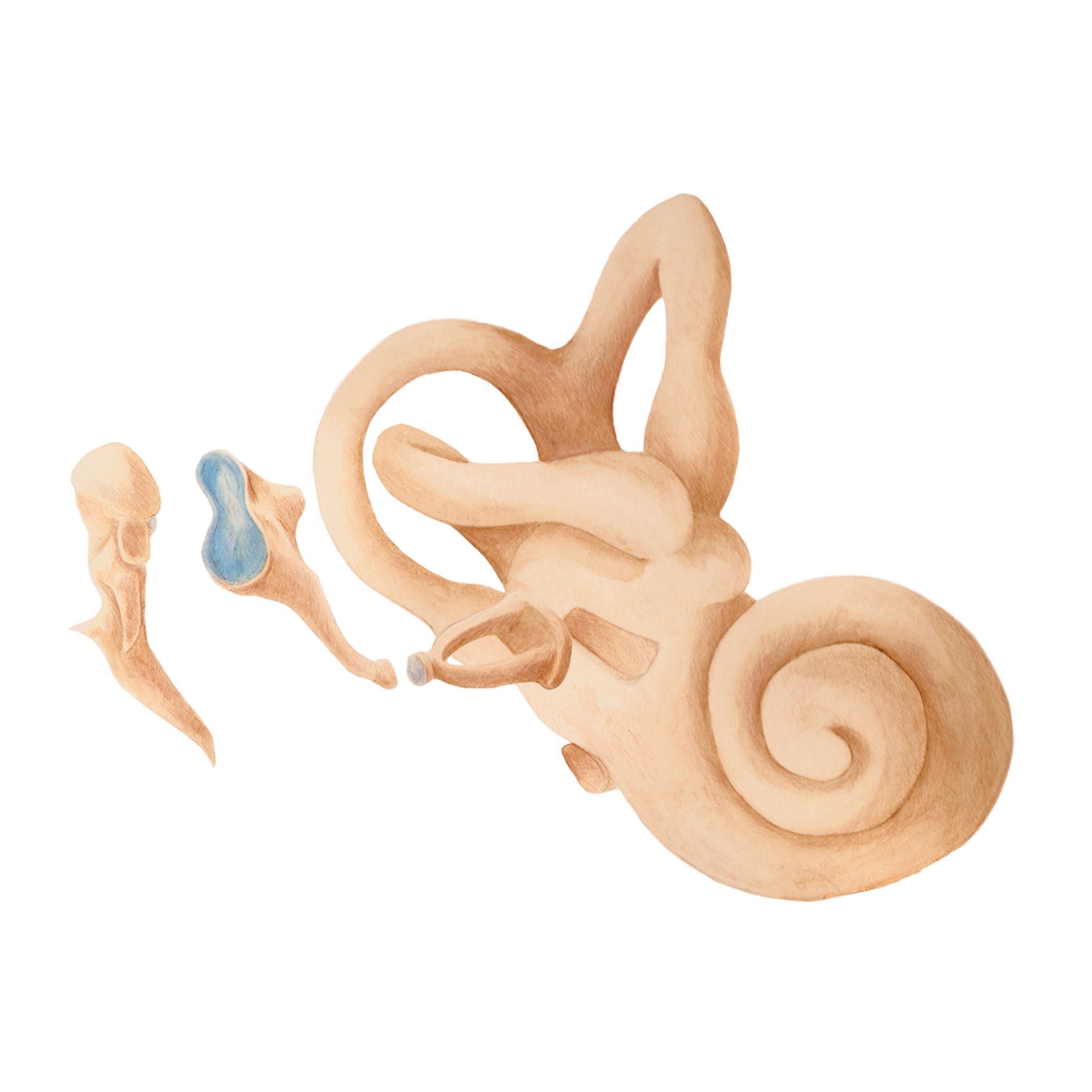
The ear (a) is a sensory organ that, in mammals, is responsible not only for hearing but also for balance. Sound waves are captured by the eardrum and transferred through the middle ear by the ossicular chain, consisting of the malleus (b), incus (c), and stapes (d). The stapes acts on the labyrinth or inner ear (e). The labyrinth is divided into two parts with distinct functions. The vestibule and semicircular canals are responsible for maintaining balance thanks to specialized cells in the ampullary crests (g) and otolithic maculae (h). The cochlea (f) is responsible for hearing thanks to the organ of Corti (i), which transforms the mechanical signal picked up by the eardrum into nerve impulses.
The idea
The idea was to create a detailed and educational illustration of the internal anatomy of the human ear, aimed at a general audience. The illustration seeks to facilitate visual understanding of the structure and function of the different parts of the ear, helping in the teaching and dissemination of topics related to hearing and balance.
The result
A precise and visually appealing scientific illustration was developed to represent the internal structures of the human ear. Each element was drawn individually using traditional techniques such as watercolor and colored pencils. The final composition was then digitally assembled, integrating all the pieces into a single work. Since anatomy is often perceived as cold, distant, and unremarkable, traditional techniques were chosen to achieve a more artisanal and warm finish, making the illustration more approachable and visually appealing.
